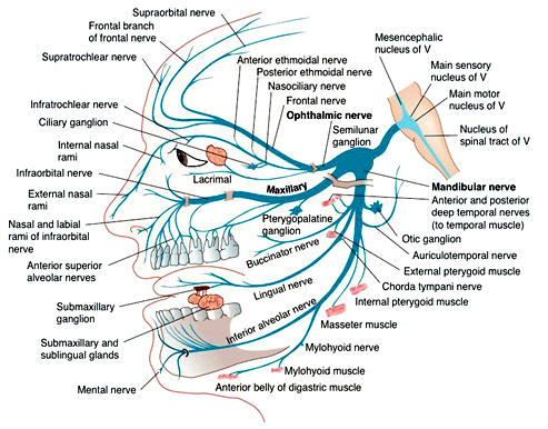
Thanks Cleo! Awesome image. Can you post the hyperlink to this? I am wanting to see more details. Any idea what the brown is over the maxillary and mandibular nerve branches? And I guess the pink is where the nerves end?
Cleo said:
You can use your pc zoom to enlarge this image or you can find over 40,000 more just by searching- trigeminal nerve images. The brown in this image under the lower teeth is jaw bone. the round darker brown (almost orange) above the lower teeth (under the tongue) and behind the eye are ganglions. The tiny pink is muscles off the tiny branches of the mandibular nerve. Those 3 main branches. ophthalmic - maxillary - mandibular, have many small nerves branching off them as shown in blue. Hope this helps??
Thanks! Over the maxillary and mandibular branches, near the semilunar ganglion is the brown I cannot figure out. I just wonder if that is where so many people get the V2 and V3 affects simultaneously.
I have a degenerative TMJ area so I think my pain comes right from that ganglion area. I also get pain above the TMJ near my temple, so it is very concentrated near that semilunar ganglion. Ug! Now it makes sense… Doesn’t solve anything, but sheds a lot of light to why I have pain there.
Thanks for sharing Cleo!
Yeah, thanks Cleo. Your first diagram has explained some of my concerns that my Neuro had just brushed over without really explaining too much. It's the first time I've seen one that detailed. :-)
